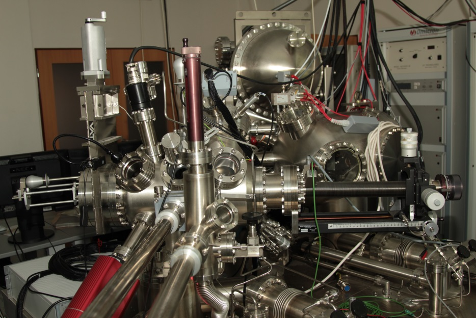
NanoESCA
NanoESCA is a new sophisticated analytical instrument designed for imaging and small spot photoemission spectroscopy (XPS) with photoemission electron microscopy (PEEM).
Excitation sources range from a high-power monochromatic Al Kα( x-ray) source to UV sources such as He-, Mercury arc- and Deuterium lamps allow to work in three regimes:
- k-PEEM momentum microscope
- classical ESCA mode
- 2D imaging ESCA and PEEM modes. PEEM images a surface of a sample with lateral resolution < 50 nm and with X-ray source better than 500 nm and energy resolution approx. 0.3 eV. The optical column provides 2D cuts of momentum distribution of photoexcited electrons (ARPES) with energy resolution approx. 0.1 eV and angle of acceptance better than ± 0.1 Å-1.
AXIS Supra

The Axis Supra photoelectron spectrometer is equipped by an Ar cluster ion source. The ion source is capable to generate Arn+ clusters consisting of hundreds or thousands of Ar atoms. As the energy of the cluster ion is shared by all atoms in the cluster the energy per Ar atom can be as low as a few eV. At this energy cluster ions only sputtered material from the near surface region leaving the subsurface layer unmodified and undamaged. Therefore the cluster ion source can be used for depth profiling of multi layer organic and inorganic materials. The cluster ion source can also be operated in standard monoatomic Ar+ mode.
XPS spectroscopy and imaging results can be complemented by the ion scattering spectroscopy (ISS) technique.
